What qEEG (quantitative EEG) is, how it is done and what it can show
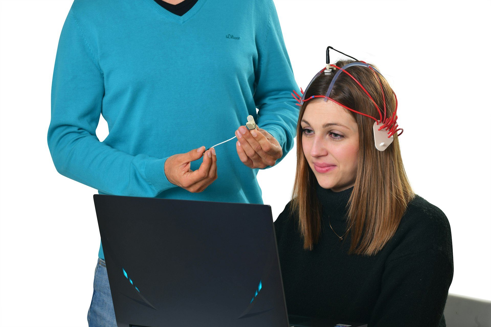
qEEG, or quantitative EEG, is an electroencephalogram analysed with the help of a computer. The brain's electrical activity is recorded with electrodes placed on the scalp, then a program turns the signal into numbers and colour maps, which it compares with the typical values for your age. The result is often called brain mapping or a brain map.
The term comes up more and more often in conversations about ADHD, anxiety or mental fatigue, and the promises that come with it vary widely. Some are accurate, others seriously stretch what a measurement can do. Below you will find how a qEEG is done, how it differs from the EEG your neurologist orders, what medical guidelines say about its limits and how much it costs at the BrainMap Institute.
What qEEG (quantitative EEG) is
The brain constantly produces tiny electrical currents, and some of them reach the surface of the head, where electrodes can pick them up. A standard EEG draws them as wavy lines, which a doctor reads by eye, page by page.
qEEG starts from exactly the same lines. The difference comes after the recording, when a program breaks the signal down into the frequency bands that have been known for a long time: delta (below 4 Hz), theta (between 4 and 8 Hz), alpha (between 8 and 13 Hz) and beta (above 13 Hz). For each electrode, it calculates how strong each band is, how the areas communicate with one another and how symmetrically the two hemispheres work.
The numbers are then compared with a normative database, that is, with the values measured in a large group of people of the same age. Deviations from the average are coloured in on a map of the head, which is where the name brain mapping comes from. A red or blue area simply means that the value there differs statistically from the average.
The method is not new. The American Psychiatric Association published a report on it as early as 1991, and the databases have grown considerably since then.
The difference between EEG and qEEG
Both tests begin in the same way, with electrodes on the scalp. They only part ways in how the signal is read and in the question they try to answer.
| Standard EEG | qEEG (quantitative EEG) | |
|---|---|---|
| How it is read | Visually, by a doctor trained in EEG | By calculation across frequencies, then interpreted by a specialist |
| What it looks for | Abnormal discharges, seizures, slowing of the trace | Patterns of power and connectivity, compared with a normative database |
| What you receive | A written report on the trace | Colour maps, scores and an interpreted report |
| Where it is used | Epilepsy, intensive care monitoring, sleep studies | Complementary assessment and research, alongside the clinical examination |
A standard EEG answers the question of whether there is anything abnormal in the trace. qEEG tries to describe how your brain works compared with that of other people your age. That is why a qEEG does not replace the EEG a neurologist orders when epilepsy is suspected, and a routine EEG was never designed to describe an attention profile.
In 1997, the joint guideline of the American Academy of Neurology and the American Clinical Neurophysiology Society required brain mapping to be used only as an addition to the standard reading of the EEG, and only by doctors highly trained in EEG. The document has since been retired. The core idea has stayed in the literature, however, and the map is read together with the trace and the patient's history.
How a qEEG assessment (brain mapping) is done
At the BrainMap Institute the assessment has a few simple steps, and the part where you sit in a chair with the cap on your head takes less time than a coffee break.
The part at the clinic takes around 20 minutes
- You have a free 15-minute phone call with a specialist to see together whether the assessment makes sense for what is troubling you.
- You are fitted with a cap with 19 soft electrodes moistened with saline solution, and fitting it takes under 5 minutes.
- You sit for 7 minutes with your eyes closed and 7 minutes with your eyes open, relaxed and without talking.
- The data are analysed and interpreted by Dr Alina Diana Nemeș and clinical psychologist Alina Robu, within 7 working days at most.
- You receive the map, the interpretation and the recommendations by email, and you can then discuss them in a consultation lasting 30 to 60 minutes.
Why two rounds? With your eyes closed, a regular alpha rhythm appears at the back of the head, and it drops sharply as soon as you open your eyes. The way the brain moves from one state to another is information in itself, so neither round is skipped.
The cap only reads. The device sends no current and does not stimulate the brain, just as a microphone does not speak on behalf of the person holding it.
What a brain map can show and what it cannot
A qEEG map can show where slow activity is stronger than you would expect at your age, where fast rhythms are in excess or how well areas far apart from each other synchronise. For a specialist, these are useful clues when they match what the patient describes.
- A profile of electrical activity at rest, compared with an age-based normative database.
- Clues about patterns that studies have linked to attention, sleep or alertness.
- A measurable starting point you can compare yourself against after a few months.
What a qEEG does not do. A brain map does not make a diagnosis on its own and does not replace a neurological or psychiatric consultation, nor the standard EEG ordered when epilepsy is suspected. If you have seizures, fainting spells, new and very severe headaches or weakness on one side of the body, see a doctor first.
This caution is backed by the literature. The American Psychiatric Association's 1991 report found qEEG useful mainly for the abnormal slow waves seen in delirium, dementia or intoxication, and noted that its role in diagnosing depression or schizophrenia had not yet been established. The American neurologists' 1997 guideline considered the method still at the research stage for attention and learning disorders, depression and mild head injuries.
Things have moved on since then, but more slowly than the advertising suggests. A 2006 report by the American Neuropsychiatric Association recommended cautious use of qEEG in children with attention and learning disorders, as well as in adults with mood disorders or dementia. In 2021, the American Clinical Neurophysiology Society guideline concluded that the evidence does not support qEEG for diagnosing concussion.
The practical conclusion is simple. The map is one piece of a bigger picture, which includes your history, the clinical examination and other tests where needed. It can also guide a brain training programme such as neurofeedback, if the specialist considers that it makes sense in your case.
What studies say about qEEG in ADHD
ADHD is where qEEG has raised the most hopes. Many studies have reported a higher ratio between slow theta waves and fast beta waves in children with ADHD.
In 2013, the US Food and Drug Administration (FDA) allowed a device called NEBA to be marketed, which measures this ratio at a single electrode on the top of the head. The approved indication is strict. The device is used in patients aged 6 to 17 and only together with the clinician's assessment, as support for a diagnosis that has already been weighed up, not as a stand-alone test.
Also in 2013, a meta-analysis of 9 studies, with 1,253 children and adolescents with ADHD and 517 without, reached an uncomfortable conclusion. The difference between the groups was shrinking from one year to the next, and the authors wrote that a raised theta/beta ratio cannot be considered a reliable diagnostic measure. A subgroup of children does have clearly different values, however, and in them the measure might say something about how things develop.
A 2018 analysis showed that fixed frequency bands can be misleading, especially in children, whose brains mature quickly. This leads to a common-sense rule. An ADHD diagnosis is made by a psychiatrist or child psychiatrist, based on the clinical interview and the diagnostic criteria, and the map can complete the picture. If you are wondering what it can add for an adult, we have written separately about qEEG assessment in adult ADHD.
EEG and qEEG in children
An electroencephalogram can be done at any age, because it only listens to the signal. With children the challenge is a different one. A 5-year-old struggles to sit for 7 minutes with their eyes closed, and every blink, scratch or smile leaves traces in the signal.
These segments are called artefacts and are removed before the calculation. When too few clean seconds are left, the result becomes less reliable, so a calm recording matters more than a long one.
There is one more important detail. A child's brain changes its rhythms as it grows, and slow waves are more present at younger ages. That is why the comparison is always made with children of the same age, never with adults. The device used at the institute can be used from the age of 2, and whether the assessment makes sense at your child's age is worth clarifying in the initial conversation.
A few things help a lot. Explain to your child beforehand, in simple words, what is going to happen, for example that they will wear a soft, slightly damp cap that only listens to how the brain works. Choose a time of day when they are usually rested and have eaten.
How to prepare for a qEEG
Preparation takes little time, but it directly affects the quality of the signal.
- Wash your hair the evening before or that morning and don't use conditioner, gel, hairspray or oil, because they prevent the electrodes from making contact with the scalp.
- Sleep as much as you usually do, because a sleepless night changes the brain's rhythms and can load the map with drowsiness.
- Eat normally and arrive without rushing, so that you have a few quiet minutes before the recording.
- When you book, mention any medicines or supplements you take, because some of them visibly change the EEG trace.
- Keep taking your treatment exactly as your doctor prescribed and change anything only with their agreement, never to make the map come out “cleaner”.
How long a qEEG takes and how much it costs
The recording itself takes 14 minutes, and with fitting and removing the cap you spend around 20 minutes at the clinic. The results arrive by email within 7 working days at most, because the map is read by people, not just coloured in by a program. We have described each stage in the article on how long a brain map takes.
The BrainMap assessment costs 2,000 lei including VAT and is paid once. The price includes the qEEG scan, the interpretation and the recommendations from the institute's founders. You will find the full list of what it includes, along with our other fees, on the pricing page.
If you want to know what the map can show for a specific problem, we have a dedicated page on anxiety and qEEG assessment. Everything you receive with the assessment is described on the BrainMap, the brain map page. Anyone who wants a more technical read on normative databases and connectivity can download the free guide to clinical qEEG.
Do you want to understand what is happening in your brain?
Book a call with the BrainMap Institute’s specialists and find out how a BrainMap and brain training can help you.
The qEEG assessment (brain mapping) is carried out at our centers in Bucharest, Cluj-Napoca, Timișoara, Iași, Brașov, Constanța, Târgu Mureș, Bacău and Craiova. Prices are listed on our pricing page.
Frequently asked questions about qEEG
What is qEEG?
qEEG, or quantitative EEG, is an electroencephalogram processed by computer. The brain's electrical activity is recorded with electrodes on the scalp, then the signal is split into frequencies and compared with the typical values for your age. The result is shown as a map of the brain, also called brain mapping.
What is the difference between EEG and qEEG?
Both start from the same recording. With a standard EEG, the doctor reads the trace by eye and looks for abnormal discharges, for example in epilepsy. With qEEG, the signal is measured mathematically and compared with a normative database, to describe how your brain works compared with other people of the same age.
Does qEEG hurt?
qEEG doesn't hurt at all. The cap has soft, damp electrodes that only read the activity your brain produces anyway. The device sends no current and uses no needles, and the only effort is to sit still for the 14 minutes.
How much does a qEEG cost?
At the BrainMap Institute, the qEEG assessment costs 2,000 lei including VAT, in a single payment. The price covers the scan, the interpretation and the recommendations, and the results arrive within 7 working days at most. All the details are on the pricing page.
How long does brain mapping take?
The recording takes 14 minutes, half with your eyes closed and half with them open, and with fitting the cap you spend about 20 minutes at the clinic. The results, with the interpretation and recommendations, arrive by email within 7 working days at most.
Can qEEG be done in children?
Yes, an EEG can be done at any age, and the device used at the institute can be used from the age of 2. The child's result is compared with reference values for their age, because brain rhythms change a lot during childhood. The hardest part is getting the little one to sit still for the 14 minutes.
Can qEEG diagnose ADHD?
Not on its own, and the studies show this too. A 2013 meta-analysis found that the theta/beta ratio is not a reliable diagnostic measure for ADHD, and the FDA-cleared device for this ratio is only used together with a clinical assessment. The diagnosis is made by a psychiatrist, and the map can complete the picture.
Sources
- American Psychiatric Association Task Force on Quantitative Electrophysiological Assessment. Quantitative electroencephalography: a report on the present state of computerized EEG techniques. The American Journal of Psychiatry, 1991. [online] Available at: pubmed.ncbi.nlm.nih.gov/2053652.
- Nuwer, M. Assessment of digital EEG, quantitative EEG, and EEG brain mapping: report of the American Academy of Neurology and the American Clinical Neurophysiology Society. Neurology, 1997 (guideline later retired). [online] Available at: pubmed.ncbi.nlm.nih.gov/9222209.
- Coburn, K.L., Lauterbach, E.C., Boutros, N.N., Black, K.J., Arciniegas, D.B., Coffey, C.E. The value of quantitative electroencephalography in clinical psychiatry: a report by the Committee on Research of the American Neuropsychiatric Association. The Journal of Neuropsychiatry and Clinical Neurosciences, 2006. [online] Available at: pubmed.ncbi.nlm.nih.gov/17135374.
- Tenney, J.R., Gloss, D., Arya, R., Kaplan, P.W., Lesser, R., Sexton, V., Nuwer, M. Practice Guideline: Use of Quantitative EEG for the Diagnosis of Mild Traumatic Brain Injury: Report of the Guideline Committee of the American Clinical Neurophysiology Society. Journal of Clinical Neurophysiology, 2021. [online] Available at: pubmed.ncbi.nlm.nih.gov/34038930.
- Arns, M., Conners, C.K., Kraemer, H.C. A decade of EEG Theta/Beta Ratio Research in ADHD: a meta-analysis. Journal of Attention Disorders, 2013. [online] Available at: pubmed.ncbi.nlm.nih.gov/23086616.
- Saad, J.F., Kohn, M.R., Clarke, S., Lagopoulos, J., Hermens, D.F. Is the Theta/Beta EEG Marker for ADHD Inherently Flawed?. Journal of Attention Disorders, 2018. [online] Available at: pubmed.ncbi.nlm.nih.gov/25823742.
- U.S. Food and Drug Administration. De Novo Classification Request for Neuropsychiatric EEG-Based Assessment Aid for ADHD (NEBA) System, K112711. accessdata.fda.gov, 2013. [online] Available at: www.accessdata.fda.gov/cdrh_docs/reviews/K112711.pdf.